Radiology reports immediately after the exam of CBCT, which is before surgery. It helps to reduce liability that may present to the implanting clinician. On the other hand, formal radiology reports accountability may be gained from several sources. It is preferably from the proper qualified as well as board certified maxillofacial radiologist. There are several types of radiology services that are present in the current day’s healthcare plan (de Baere, 2015). CT scan or computed axial tomography is one of the types that deal with a non-invasive medical test. It utilises special x-ray equipment with different types as well as sophisticated systems to generate several images or pictures of the inside of the body.
On the other hand, CT lung cancer screening deals with low dose CT scan of the lungs that are used to identify lung cancer. Also, fluoroscopy deals with a medical test where a constant x-ray beam is passed across the body. However, magnetic resonance imagining scan is essential for the procedure where it is required to use a non-invasive medical test or examination that utilises a large magnet as well as a computer taking an image of the inside body. Ultrasound named as sonography we considered as a medical test that uses high-frequency sound waves. A computer creates images of the blood vessels, tissues as well as organs (Anzidei et al., 2015). The virtual imaging process utilises CT scanning as well as advanced computer hardware to generate 2d as well as 3d images of the colon, which can be viewed on the video screen. X-rays is one of the most popular techniques that uses invisible electromagnetic energy beams to generate the images of internal tissues, bones as well as organs on a film.
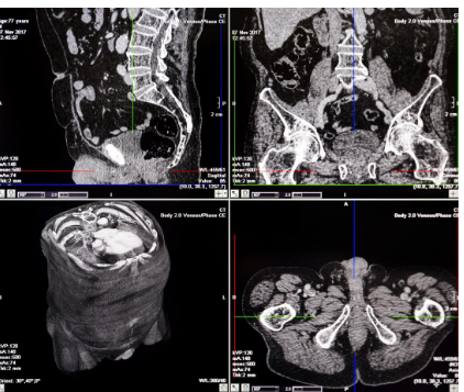
Figure 1: CT scan report
(Source: Charest-Morin et al., 2016, p.141)
In the present study, Mr A Tomy has been selected as a case study person. He is 55 years old and assaulted during walking home from a local game. After the injury, he was senseless. However, he regained it when in the ambulance (Charest-Morin et al., 2016). It has been found that significant soft tissue swelling, as well as bleeding to the left eye and side of the head, found apparent. In this situation, it is essential to perform radiology for the patient at first so that the spot and density of the injury can be located appropriately.
In this situation, the CT scan can be the most suitable radiology that would be helpful for the patient to identify the exact spot as well as the density of the injury. Molvar and Glaenzer (2016) stated that the radiology helps in diagnosing the muscle as well as bone disorders like bone as well as features. The process can detect the pinpoint of the specific location of the injury and blood clot. It also allows the doctor to check the depth of the damage inside the eye as well as the head. It utilises a combination of X-rays as well as a computer to develop pictures of the organs, bones as well as different tissues.
The CT scan process utilises a narrow x-ray beam that can circle the parts of the body. It also provides a series of images of the body. The procedure is generally developed to generate a detailed picture of the organs and blood vessels (Cheshire et al., 2016). For instance, the surgeon may utilise the scan for preparing an operation. CT scan can be useful to detect the bone injury as well as joint problems such as complicated bone features as well as tumours. In addition, they can assist in locating the injuries as well as blooding like it is caused by accident. Doctors use for guiding the treatment plans as well as processes like biopsies and surgeries as well as radiation therapy (Cheshire et al., 2018). In addition, it helps to compare finding out whether specific treatments are working correctly. The scans of the injury places over time can show if the responding chemotherapy or radiation.
With the help of
CT scan, the solid substances such as bones injury of Mr Tomy can be easily
seen. However, the soft tissues do not show up. To assist in the process, it is
required for a special dye named as contrast material (Molvar & Glaenzer, 2016). The x-rays can be blocked
as well as appear on the scan. It helps to highlight the blood vessel, organs
as well as different structures. Hence, it is essential to select the best
possible and suitable radiology for the patient so that proper treatment can be
performed and the patient recovers quickly.
References
Anzidei, M., Lucatelli, P., Napoli, A., Jens, S., Saba, L., Cartocci, G., Sedati, P., d’Adamo, A. & Catalano, C., 2015. CT angiography & magnetic resonance angiography findings after surgical & interventional radiology treatment of peripheral arterial obstructive disease. Journal of cardiovascular computed tomography, 9(3), pp.165-182.
Charest-Morin, R., Boriani, S., Fisher, C.G., Patel, S.R., Kawahara, N., Mendel, E., Bettegowda, C. & Rhines, L.D., 2016. Benign tumours of the spine: has new chemotherapy & interventional radiology changed the treatment paradigm?. Spine, 41, pp.S178-S185.
Cheshire, S.C., Board, R.E., Lewis, A.R., Gudur, L.D. & Dobson, M.J., 2018. Pembrolizumab-induced sarcoid-like reactions during treatment of metastatic melanoma. Radiology, 289(2), pp.564-567.
de Baere, T., Deschamps, F., Tselikas, L., Ducreux, M., Planchard, D., Pearson, E., Berdelou, A., Leboulleux, S., Elias, D. & Baudin, E., 2015. GEP-NETS update: Interventional radiology: role in the treatment of liver metastases from GEP-NETs. European journal of endocrinology, 172(4), pp.R151-R166.
Molvar, C. & Glaenzer, B., 2016, December. Choledocholithiasis: evaluation, treatment, & outcomes. In Seminars in interventional radiology (Vol. 33, No. 04, pp. 268-276). Thieme Medical Publishers.